Basal Ganglia and Parkinson’s Disease: How are they Connected?
The Basal Ganglia refers to a group of structures located beneath the cortex of the brain called “subcortical nuclei”. With Parkinson’s Disease, there is dysfunction within the basal ganglia. In this blog post, we will discuss the anatomy of the basal ganglia and the pathophysiology of Parkinson’s Disease.
Functions of the Basal Ganglia:
The Basal Ganglia have many responsibilities including:
Aiding in motor learning (learning new movements/actions)
Executive functions (such as decision making, planning etc)
Emotional behaviors
Addictive behaviors
Anatomy:

The subcortical nuclei that together make up the basal ganglia include:
Striatum – This is the largest structure of the Basal Ganglia. It is comprised of two main divisions:
Dorsal Striatum – This consists of the caudate nucleus and the putamen. The dorsal striatum is involved in motor movement and executive functions such as decision making and planning.
Ventral Striatum – This consists of the nucleus accumbens and the olfactory tubercle. The ventral striatum is responsible for limbic functions like reward and aversion.
Globus Pallidus – helps to regulate movement especially subconscious movement
Subthalamic Nucleus – this structure is involved in suppressing unwanted movements
Substantia Nigra – Substantia Nigra means “black substance” in latin and it got this name because a dopamine precursor in this area of the brain has a black pigment causing this structure to be a black in color. This is the structure in the basal ganglia that produces dopamine.
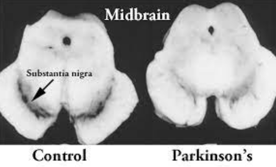
This image shows how there is a dark pigment to the substantia nigra in healthy controls, however, with the loss of dopamine in those with PD, you can see how the dark pigment is lost.
Parkinson’s Disease Pathophysiology:
Appropriate functioning of the basal ganglia system requires dopamine to be released. With Parkinson’s Disease, the dopaminergic neurons in the substantia nigra degenerate progressively which leads to severe dopamine deficiency and the cardinal features of Parkinson’s Disease (tremor, bradykinesia, rigidity, and postural instability).
At the point that clinical signs of Parkinson’s Disease are evident, about 80% of the striatal dopamine and 50% of the nigral neurons are lost. The neurons that are still alive, often have misfolded proteins such as alpha-synuclein which causes circular shaped aggregates called Lewy Bodies. Lewy bodies are not just in the substantia nigra, but they can be found in the cortex of the brain, the amygdala (which houses our emotions), and the vagal nucleus (a cranial nerve that regulates and calms internal organ functions such as digestion, heart rate, respiratory rate). Therefore, Lewy Bodies in these non-motor areas could account for many of the non-motor symptoms that are found in Parkinson’s Disease.
The Basal Ganglia is involved in pathways that are inhibitory or excitatory so they can cause more motion or discourage motion. The structures of the basal ganglia can be broadly categorized as input nuclei (that receive input), output nuclei (that send out information) and intrinsic nuclei (that relay information between the input and output nuclei). With Parkinson’s, there is a lack of dopamine which leads to too much inhibition. This is what causes bradykinesia (slowed movements), hypokinesia (small movements) and many of the other common symptoms of Parkinson’s Disease.
Now you hopefully have more of an understanding of the role of the Basal Ganglia and what happens with Parkinson’s Disease. There is so much that can be done to help with symptom management such as medication management and exercise and more and more research is showing how exercise can help delay progression of Parkinson’s Disease. For more on this, check out our other blog posts - especially the one on aerobic exercise for PD.
Sources:
Lanciego et al, 2012. Functional neuroanatomy of the basal ganglia. CSH Perspectives in Medicine.
Samii et al, 2004. Parkinson’s Disease. The Lancet 363: 1783-1793.